First part: functional anatomy of the knee ligament
To fully understand knee laxity and relate it to a specific ligament lesion, it is essential to first know the normal functioning of the knee, which requires some essential prior knowledge:
- anatomical of course
- kinematics of joint movement mechanisms
- biomechanics of the forces and constraints exerted on the knee
- on the anatomo-functional correlation between the perfectly invisible anatomical ligament lesion and its functional repercussion, laxity, clinically testable.
The impossibility of having direct visual contact with the anatomical reality of a ligament lesion makes clinical examination essential in the diagnosis of ligament lesions of the knee; it is then possible to get a precise idea of the lesions through the examination signs.

1- Functional anatomy
The knee is a complex joint system that includes 2 distinct joints:
1- the femoro-patellar joint between the femoral trochlea and the internal face of the patella with its own anatomy and physiology and without interest for the knowledge of the ligamentous knee.
2- the two internal and external femoro-tibial joints (medial and lateral) in which laxity is expressed. They bring into contact the 2 internal and external femoral condyles (CE and CI) and the 2 internal and external tibial plateaus (PTI and PTE). These 2 femoro-tibial compartments are fundamentally different both in their design and in their operation:

The two femoro-tibial compartments

Congruent internal compartment: convex condyle, concave tibial plateau

Incongruent external compartment: condyle and tibial plateau are convex
- the internal femoro-tibial compartment is synonymous with stability. It ensures effective joint coaptation via a powerful ligament system and suitable bone complementarity between the convexity of the internal condylar pulley and the concavity of the tibial glenoid.
- on the other hand, the outer femoro-tibial compartment is designed for mobility. Articular coaptation in this lateral compartment is less because of the low tension of its capsulo-ligamentous apparatus and its articular incongruence with 1 CE twisted on its sagittal axis in the shape of a convex bean and an equally convex PTE.

Intercondylar notch in which the thorny massif intertwines
Functionally this double F/T joint is described as a bi-condylar joint. In reality, it functions like a trochlear joint modified by the spinous mass which allows 2 degrees of freedom: flexion/extension and rotation.
1 2 3


Bicondylar (1), trochlear (2) and modified trochlear (3) type joint, the latter corresponding to the functional reality of the knee
- in the sagittal plane, the movement of flexion/extension (F/E) takes place by rolling-sliding of the condyles on the tibial surfaces, while avoiding early subluxation of the femur and with a large amplitude of movement of the order of 150°.

- in the horizontal plane , the incomplete embryological genesis of the tibial trochlear crest of the massif des épines allows rotational movements (R) whose amplitude varies according to the flexion of the knee: 29° from R to 60° from F and 26° to 90° from F.


In the horizontal plane, rotational movements of the condyles on the tibial plateaus from 29° to 60° of Flexion and from 26° to 90° of F.
- in the frontal plane, there is no physiological movement except a small mobility in varus corresponding to a minimal adaptation to the constraints of Varus/Valgus.

No possibility of movement in the frontal plane
This double F/T joint has sagittal axes of movement of F/E and horizontal in R which are perpendicular and the place of geometric interception of these axes, defines the articular center of the knee (AGC) corresponding to the point of application of the multidirectional forces which are exerted on this CAG, which merges with the solid mass of the tibial spines and thus with the ligaments of the central pivot. We can therefore say that the CAG = the central pivot ligament.

The knee joint center (AGC) corresponds to the interception of the sagittal axes of F/E and horizontal of R
2- Kinematics
Whoever says articulation, necessarily says movement and therefore the presence of a muscular motor which will act on the axes of the CAG. This driving force will be of variable intensity, depending on the power of the muscular contraction. It will have a fixed direction, parallel to the longitudinal axis of the muscle fibers which will depend on its point of application with respect to the CAG. If the point of application (PA) of the driving force is close to the axis of flexion, there will be no sagittal movement and the muscle will only be rotator. If the PA is close to the axis of rotation, the movement will be sagittal in F or in E. In the other hypotheses if the PA acts on the 2 axes, there will be both F and R.

The driving force has a fixed direction // at the axis of the muscle fibers and a point of application: PA, located differently with respect to the CAG and the sagittal and horizontal axes
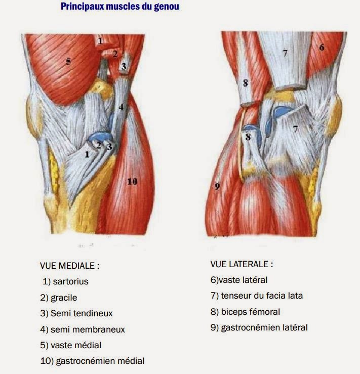
For the knee, the muscular motor is made up of the peri-articular muscles:
- the Quadriceps is extensor and a little internal rotator.
- the Twins are powerful flexors and not at all rotators.
- the lateral muscular stays (Tensor du facia lata and Biceps femoris) are only flexors + external rotators.
- the medial shrouds (crow's feet muscles) are only internal flexors and rotators.
Disruptive muscle forces
Alas, this muscular motor has at the same time a serious drawback, that of generating disturbing forces which no longer act on the axes of the CAG but directly on the bone structures, in this case for the knee on the tibia. If we take the example of the Quadriceps, it will exert a disturbing force on the tibia in the fully extended position and up to 60° of flexion, first in the anterior drawer then in the posterior drawer. For the twin muscles of the calf, they will pull the condyles on which they are inserted backwards and therefore exert a deleterious disturbing constraint in the anterior drawer, and this regardless of the degree of flexion of the knee.
3- Stabilizing action of the menisci

The 2 menisci are triangular fibrocartilages at the cut which are interposed between the condyles and the tibial plateaus. The internal meniscus (MI) is open at C, the external meniscus (ME) is closed at O. They are 4cm long, while their width and thickness do not exceed one cm. They are adherent to the capsule by their circumferential edge (much more the MI) and are fixed at the level of their anterior and posterior horns. They act as a wedge and improve the F/T congruence while distributing the stresses. They have a dynamic effect through their posterior connections with the antero-internal (PAPI) and postero-external (PAPE) angle points, with the semi-membranosus for the MI and the popliteal muscle for the ME. Knee in flexion they move forward (by 6cm for the ME) and knee in extension they move back.

displacement of the menisci during knee flexion
4- Stabilizing action of the various knee ligaments and fibrous formations
The ligaments of the knee, like all ligaments, are elastic elements composed of collagen fibers which make them restoring forces whose stabilizing action will be proportional to their state of tension. There is a nonlinear relationship between the restoring force developed by a ligament and its elongation. The 2 variables are not proportional: at the beginning of the solicitation of a ligament, the restoring force is less, then once the fibers have been recruited, the ligament undergoes a phase of elastic deformation with elongation without damage to the ligament. If the constraint continues, there will be partial, then total rupture without parallelism with the restoring force developed by the ligament which does not increase.

Center pivot stabilizing action
Because it merges with the CAG, the stabilizing action of the central pivot: anterior cruciate ligament (ACL) + posterior cruciate ligament (PCL) is decisive.
The ACL is inserted on the tibia anteriorly on a triangular surface of 3 cm2 (prespinal surface) and externally on the condyle on a surface of 2 cm2 positioned sagittal.
It has a twisted appearance and is made up of 2 bundles: 1 main anteromedial bundle, larger and longer than the other bundles, it controls anterior translation. The more slender posterolateral bundle is positioned more vertically and controls internal rotation. Some authors even describe an intermediate beam. Whatever the position of the knee, the ACL is always in tension: knee in extension, the 2 beams are vertical and tense; knee flexed, the ACL becomes horizontal, the main bundle is distended, but the PL bundle remains taut because it is vertical.
The PCL is inserted on the tibia posteriorly over an area of 0.5 cm2 (retrospinal surface) and on the medial condyle. Oriented at 45° in the 3 planes of space, it is made up of 2 bundles, a posterolateral bundle oriented vertically and a small anteromedial bundle oriented more horizontally. Unlike the ACL, the PCL horizontalizes the knee in extension and verticalizes in flexion; whatever the position of the knee, it is always more tense than the ACL.

In flexion-external rotation, the pivot is uncrossed and cannot control this external rotation
In the end , the central pivot ligament stabilizes the knee in the 3 planes of space and particularly in sagittal where the ACL controls the anterior translation and the PCL the posterior translation. In the horizontal plane the ACL wraps around the PCL and controls internal rotation. The external rotation escapes the pivot completely uncrossed and ineffective in this position. In the frontal plane, when the knee is straight, the pivot opposes the varus/valgus stresses. When the knee is flexed, the varus/valgus is controlled by the collateral ligaments LLI for valgus and LLE for varus .
Posterior fibrous formations
They correspond to a fibrous sheet placed behind the inter-condylar space and the crosses. It is continued on the sides with the condylar shells which are closely related to the tendons of the gastrocnemius, popliteal and semimembranous muscles.
They have bundles, some of which are more individualized, such as the oblique popliteal ligament which detaches from the 1/2 Membranous and the arcuate popliteal ligament which forms an arch for the Popliteal muscle. They form a functional whole with the posterior horns of the menisci which are added to them. These are the first brakes on the advancement of the tibia and therefore on the control of the external rotation of the knee in flexion.
Internal training: LLI and posterior internal: PAPI.

LLI and PAPI (posterior oblique ligt or LOP + post horn of MI + tendon and semimembranosus muscle)
The LLI is a very strong ligament, tightly adherent to the capsule, and runs from the medial condyle to the medial proximal tibial metaphysis. It opposes constraints in valgus stretched knee with the pivot and flexed knee, alone. With the PAPI it controls the RE by slowing down the advance of the ITP and also the anterior translation.
THE PAPI is a very resistant formation, made up of the posterior oblique ligament (LOP), the posterior horn of the MI and the tendon of the semimembranous muscle which is the active element of the PAPI. It additionally controls the posterior drawer, internal rotation and valgus in a position close to extension. The Dupont test in external rotation tests the PAPI.
External training: LLE and postero-external: PAPE.
External training: LLE and postero-external: PAPE.
The stretched LLE from the external condylar tubercle to the head of the fibula does not adhere to the capsule. It alone controls the varus when the knee is flexed; when the knee is stretched it participates in its control with the PCL.
PAPE is less resistant than PAPI. It is made up of the posterior horn of the external meniscus, the external condylar shell, the tendon and popliteal muscle which is its active element, the fibrous popliteal complex and an arcade in its lower part (arcuate popliteal ligament) for the main tendon of the popliteal muscle.
It prevents recoil of the external tibial plateau and must be repaired in the event of injury. It essentially controls posterior translation, external rotation and varus, knee at 30° flexion. The Reverse Pivot Schift test of Jacob and the big toe test are positive in case of lesion of the PAPE.
LLE and PAPE stabilize external rotation, limit anterior translation and secondarily internal rotation. They have a moderate anti-varus role.

The antero-lateral formations
They correspond to Kaplan's fibers, fibrous band, pearly and resistant, described by the French surgeon Segond in an article published in 1879. This fibrous band is also described under the term of antero-lateral ligament (ALL) or ilio tibial tract of the anglo-axons. It participates in the control of internal rotation and can be injured in the event of excessive torsion of the knee in internal rotation and take away a bone fragment at their tibial insertion behind Gerdy's tubercle, known as Segond's fracture. The presence of a fracture of Segond necessarily indicates a rupture of the ACL, thehe unmooring of the antero-external ligament is always preceded by that of the ACL, so that the presence of a bony pellet carrying the external tibial margin is pathognomonic for the rupture of the ACL.


Segond's fracture = disinsertion of Kaplan's fibers
In the end
The different central and peripheral ligament elements make up a stabilizing physiological unit whose different elements partially supplement each other and act as primary or secondary brakes.
The different central and peripheral ligament elements make up a stabilizing physiological unit whose different elements partially supplement each other and act as primary or secondary brakes.
In sagittal, the pivot is the primary brake of the anterior and posterior drawer in extension and in flexion with the LLI and the PAPI for the anterior drawer and the LLE and the PAPE for the posterior drawer. In flexion and internal rotation, PAPI and LLI replace the PCL.
In frontal the primary valgus brake is the LLI and in varus the primary brake is the PAPE.
In rotation, the main brake on internal rotation is the pivot: ACL + PCL with maximum tension and internal and external stabilization. In external rotation, the main brake are the internal peripheral formations for the medial side and external for the lateral side.
l imits of the ligament stabilization system
l imits of the ligament stabilization system
The knee ligament system is no exception to the rule of any ligament system which has its own limits related to the functional rigidity of its structures.
The ACL has an elastic resistance of 60 kilos per cm2, far too little to counter the Quadriceps which can develop stress forces that can go up to 400 kilos per cm2.
The knee will therefore need a support system which at the same time will be a protection system for the ligaments and this support system will necessarily be dynamic and devolved to the various peri-articular muscles according to the precise movement needs so as not to harm. knee mobility.
This peri-articular muscular system: quadriceps, twins, crow's feet muscles and lateral stays TFL and Biceps femoris is the best articular coaptator of the knee, it ensures with the stabilizing action of the various ligaments, the biomechanical balance of the knee against disturbing forces which are thus controlled.
5- The stability of the knee in summary
Sagittal stability:
Knee in extension:
the ACL acts alone on the anterior displacement: ACL lesion = anterior translation of the tibia = Lachman to practice at several degrees of flexion between 10° and 25° always painless unless associated meniscal lesion.
Knee in flexion:
- the anterior translation is also limited by the collateral ligaments and the angle points, it is best appreciated at 70° of flexion.
Isolated lesion of the ACL = no direct anterior drawer (TAD). If TAD = ACL lesion + lateral or posterior formations, especially when the wedge represented by the MI disappears.
- the posterior translation is assessed at 90° of flexion; it is limited by the LCP and the corners.
rotational stability
The rotational movement is controlled by the Pivot: ACL when the knee is close to extension and LCP knee in flexion.
Two positions of stability: the VFE and the VRI: very often associated capsulo-ligamentary lesions will occur on an indirect forced movement or more serious if movement supported from its 2 positions of stability:

- in VFE (valgus/flexion/external rotation) the stability is predominantly active (IJ for flexion with in particular the half membranous and the popliteus for the RE and initially theoretical danger for the PAPI and the LLI in the event of a mechanism injury in VFE, which is still not verified in practice or the ACL may be injured in isolation.
- in VRI (varus/flexion/internal rotation) with activo/passive stability position by the Maissiat band and the Biceps, and danger for the PAPE and the ACL in the event of a lesional mechanism in VRI.frontal stability:
The valgus is limited by the LLI+PIVOT
- knee in extension: small valgus = lesion of the LLI = benign sprain
- great valgus = lesion of the Pivot + LLI.
The varus is limited by LLE + PIVOT (mainly LCP) + PAPE
- small physiological varus in flexion
- large varus in extension = external lesion (LLE) + posteroexternal (PAPE) + pivot
Second part: clinical evaluation of knee sprains
The knee is the relay joint of the articular chain of the lower limb and as such, it participates in the development of the movement in space of individuals and their gravitational balance during the said shift.
It will be subjected to external constraints which will compromise its intrinsic balance, and its own static and dynamic stabilizers will have to oppose these disturbing forces more or less successfully in order to lock the knee in a stable position, thus acting as energy absorbers.
Knee in extension = isolated ligament sprain


 In dynamic hyperextension, maximal contraction of the quadriceps will lead to ACL tear 3- other MLs with isolated ACL injury a/dynamic hyperflexion
 in dynamic hyperflexion, the maximum contraction of the quadriceps will cause the rupture of the ACL


 9
9
knee in flexion = multi-ligament sprain
 Maximum positions of stability that can be exceeded by an offensive external force


small recurvatum = shells
medium recurvatum = LCP
large recurvatum = LCP + posterior formations and shells.


 Pivot shift and jerk test


The knee is the relay joint of the articular chain of the lower limb and as such, it participates in the development of the movement in space of individuals and their gravitational balance during the said shift.
It will be subjected to external constraints which will compromise its intrinsic balance, and its own static and dynamic stabilizers will have to oppose these disturbing forces more or less successfully in order to lock the knee in a stable position, thus acting as energy absorbers.
Knee in extension = isolated ligament sprain
Knee in extension, static or dynamic hyper - extension are the two most frequent MLs.
In extension, the knee is in a position of static stability and its locking ensured both by the camber of its femoro-tibial articular surfaces in a position of maximum congruence and by its central and peripheral ligament system. Despite everything in this position of static stability, knee in extension, its damping potential is low and represented only by the reserves of elasticity of its capsulo-ligamentary system which is very vulnerable in the face of disturbing external stresses, especially if these are not mastered. This will then cause lesions of the stabilizing structures directly opposed to these stresses and in the first place lead to isolated lesions of the primary brakes.
1- Mechanism of injury (ML) in static hyper - extension
In static hyper - extension of the knee, foot fixed to the ground, if an injurious force is applied to the knee from front to back, the primary frenulum constituted by the posterior cruciate ligament (PCL) will rupture in isolation by displacement towards the back of the tibia under the femur (receiving a jump, tackle on the tibia in football).
Lesion mechanism in static hyperextension with isolated lesion of the PCL
2- ML in dynamic hyperextension
According to the same principle, in dynamic hyper - extension of the knee by maximum contraction of the quadriceps, such as for example a schoot in the void in football, the primary brake directly opposing the advance of the tibia under the femur is the anterior cruciate ligament (LCA) which will come to break in isolation on the easel of the inter - condylar notch.
b/ Internal rotation, knee, knee close to extension
It is a classic injury mechanism when playing sports such as football or volleyball. Upon landing a jump, the foot is blocked on the ground in internal rotation, the body pivots in the other direction, which results in internal rotation of the knee putting maximum tension on the anterior cruciate ligament, often beyond the limit of its resistance. This mechanism is often responsible for isolated rupture of the anterior cruciate ligament.
ML in VRI
3- always knee in extension: ML in varus/valgus forced by lateral or medial vulnerating force
These 2 MLs are rare and lead to lesions of the peripheral primary brakes, the external and internal collateral ligaments (LLE and LLI).9ML in forced valgus (LLI rupture) and ML in forced varus (LLE rupture)
knee in flexion = multi-ligament sprain
In bending, the situations are completely different.
Flexion lifts the static lock and frees the rotation of the knee, so that external rotation and varus as well as internal rotation and valgus limit each other and lock the knee thanks to the active control of the periarticular musculature which then behaves as a stabilizer dynamic.
Unlike static locking, this dynamic locking has a high damping potential and the peri-articular musculature of the knee has the capacity to damp external traumatic energy by adapting the position of stability according to the direction of the disturbing force, either in valgus - external rotation (VFE), or in varus - internal rotation (VRI).
A bent knee sprain therefore always corresponds to exceeding a position of stability in VRI or VFE and is accompanied by multi-ligament lesions, unlike the isolated lesions of sprains in extension.
1- VFE is the most frequent ML.
ML in VFE
- if the vulnerating force in valgus is predominant, there is immediate danger for the LLI, then the PAPI, then the ACL and if the constraint continues, the PCL.
- if the wounding force in RE is predominant, the immediate danger is for the PAPI, then the LLI, then the ACL. If the stress in RE continues, the lesion will extend to the PAPE, while the PCL, serving as a pivot, remains intact.
In VFE, the lesional association LLI + PAPI + ACL is frequent and produces the classic internal lesional triad.
ML in VFE, hemarthrosis in less than 4 hours, internal triad with ecchymosis
2- ML in VRI , there is immediate danger for the PAPE, then the LLE, then due to the excessive screwing of the central ligamentous pivot, the ACL, then the LCP, then the PAPI if the constraint continues.
In VRI, the lesional association PAPE + LLE + ACL = classic external triad. If LCP + PAPE = internal pentad. ML in VRI
Hyper-laxity of the knee
A ligament is an elastic structure, each ligament having a basic laxity and an elongation potential of its own. This baseline laxity is increased bilaterally in hyperlax subjects. Knee joint laxity is therefore a physiological phenomenon and the expression of reciprocal mobility between the femoral and tibial articular surfaces.Conversely, hyperlaxity is a unilateral pathological increase in baseline laxity, a consequence of ligament injury, which can be compensated by other healthy ligament structures, as well as muscle coaptation forces.
For each knee injury, there is therefore a well-defined type of hyper-laxity:
- an isolated lesion of the central pivot leads to pure hyper-laxity with exaggerated but symmetrical displacement of the 2 tibial plateaus forwards if the ACL is torn or backwards if it is the PCL.
- an isolated lesion of the collateral ligaments with an intact pivot leads to internal or external peripheral laxity.
- an associated lesion of the pivot and an internal or external peripheral structure leads to mixed hyper laxity with asymmetrical translation of the tibial plateaus and rotation.
Instability
In the event of a major ligament lesion, the compensation mechanisms are no longer sufficient and the knee joint center (AGC) will be destabilized and knee instability will set in. This instability, unlike hyper-laxity, is always poorly tolerated and clinically manifests as slipping away or feelings of repeated dislocation.
This instability can be present from the outset or occur secondarily, the overload of the remaining healthy ligaments, inducing their progressive distension and by so much ending up altering their stabilizing function.
Unfavorable factors
The prognosis of rotational hyper-laxity due to the greater overload of healthy elements is much worse than that of translational hyper-laxity.
Similarly, external hyper-laxities are less well supported than internal ones given the lesser structural stability of the external compartment.
The genu-varum is also an unfavorable factor, due to its impact on the external ligament structures whose reserves of functional elasticity are diminished.
The genu-recurvatum in the same way distends the posterior capsulo-ligamentous structures as well as the ACL.
Clinical diagnosis of hyper-laxity and instability of knee ligament origin
1- Sagittal hyper-laxity in anterior translation
They are highlighted by the various drawer maneuvers and are positive when there is an abnormal translation of the tibia relative to the femur, either forwards or backwards.
Knee bent at 90°
TAD = direct anterior drawer in neutral rotation, first puts the ACL in tension, then the internal peripheral structures, then the external ones.

TAD (direct anterior drawer)
TAD from 3 to 5 mm = isolated ACL.
TAD from 5 to 10 mm = ACL+ LLI or LLE.
TAD > 10 mm = ACL+LLI+PAPI+POPE
TARI = anterior drawer in internal rotation, puts tension on the ACL, then the external structures.
TARI from 5 to 13 mm = isolated ACL.
TARI > 13 mm = ACL + LLE + PAPE
TARE = anterior drawer in external rotation, decreases tension on the ACL and external structures; increases the tension of the internal peripheral structures. A positive TARE = hyper-laxity of the internal compartment: LLI and PAPI.
TARE from 5 to 13 mm = ACL isolated.
TARE from 13 to 19 mm = LLI and PAPI.
TARE > 19 mm = LLI + PAPI + ACL.
Knee flexed at 20° = Lachman-Trillat test
At 20° of flexion, the tensile force of the hamstring muscles which opposes anterior translation is zero, because this force is perpendicular to the anterior drawer, but also because the articular surfaces of the condyles, which are relatively flat, do not interfere more with the movement of the tibial plateaus.
The Lachman test, when it is positive, is very sensitive, even for very low amplitude anterior displacements .
Lachman from 2 to 9 mm = ACL ruptured alone.
Lachman > 10 mm = ACL + peripheral structures.
Lachman-Trillat
2- Posterior hyper-laxity
They are based on the same principle and are carried out using maneuvers this time in the posterior drawer = TP, not always easy to diagnose.TP < 5 mm = possible partial rupture.
TP between 5 and 10 mm = isolated LCP.
TP >10 mm = LCP + PAPE.
Rear drawer (TP)
1- unilateral recurvatum:Three other tests:
small recurvatum = shells
medium recurvatum = LCP
large recurvatum = LCP + posterior formations and shells.

Unilateral recurvatum
2- unilateral external hyper-rotation , stretched knee and bent knee.
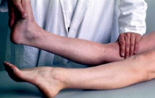
3- Godfrey's sign in dorsal decubitus, both knees bent at 90° and heels supported: unilateral recoil of the tibia, signs a rupture of the PCL.Godfrey's test (tibial metaphysis fall backwards)
3- Lateral hyper-laxity
- knee at 30° flexion, testing in forced varus and valgus:This forced varus-valgus testing at 30° explores the collateral ligaments, while the corner points are relaxed. If the test is positive, this indicates a lesion of the LLE (forced positive varus) or the LLI (forced positive valgus).
- knee in extension, if the varus-valgus test is positive, this indicates a lesion extended to the corner points and even to the central pivot.
varus/valgus testing
4- Dynamic testing of anterior or posterior instabilities.
The instabilities of the knee, whether anterior or posterior, are sought by jump tests.An anterior condylar jump can be looked for in many different ways: Lemaire tests, Mac Intosh, Jerk test, Slocum etc., but the principle is the same: in anterior instability following a rupture of the ACL, the PTE (external tibial plateau) is subluxed forward; if the knee is gradually flexed, towards 30° of flexion, the subluxation is reduced and is accompanied by a jerk perfectly identifiable by the examiner's palpating hand placed at the height of the PTE.
anterior condylar jump
If we take Mac Intosch's Pivot-schift as an example: the start of the test is carried out with the leg stretched, the foot kept in internal rotation by the mobilizing hand, the palpating hand is placed on the external tibial plateau so as to print a slight valgus on the tibia. The knee is gradually flexed and if the knee is stable, the external condyle slides smoothly on the external tibial plateau; in the event of instability, around 30° of flexion, the subluxation of the PTE is suddenly reduced and the palpating hand feels a jump under the fingers. This test is 98% reliable.
A positive posterior condylar jump signs the diagnosis of posterior instability and the tests are also very numerous. We will take Jacob's reverse condylar jump as an example: the subject to be examined is in dorsal decubitus, knee bent; by the simple fact of gravity, the PTE sub-luxates backwards, the examiner with his palpating hand, imparts a valgus movement on the PTE, while the distal mobilizing hand, foot positioned in external rotation ,extends gradually the knee and towards 30° of flexion, the subluxation is reduced with a characteristic noise. This test is also 98% reliable.
Jacob's posterior condylar jump
5- Others majors clinical signs in the diagnosis of a sprained knee
- questioning the injured person with analysis of the mechanism of injury (knee stretched, knee bent, anterior, lateral impact), history (often it is the first episode),
- inspection (swelling, bruising in case of capsular breach)
- palpation are also major times.
Among the immediate signs: the perception of a creak contemporaneous with the sports accident (suspicion of lesion of the pivot), the often acute pain in immediate post traumatic, functional impotence (generally it is impossible to get up alone after knee sprain), rapid swelling (in less than 4 hours, hemarthrosis sets in with a sign of the ice cube, but leaves time for the first hour to examine the knee.- questioning the injured person with analysis of the mechanism of injury (knee stretched, knee bent, anterior, lateral impact), history (often it is the first episode),
- inspection (swelling, bruising in case of capsular breach)
- palpation are also major times.
Rapid hemarthrosis in less than 4 hours
In the end, if we manage to clinically differentiate the different types of laxity, we can deduce the underlying anatomical lesion, the clinic allowing to strongly suspect the diagnosis at more than 90%.
But there are pitfalls that may prove insurmountable:- the pain can lead to muscle contractures which will make ligament testing impractical or uninterpretable.
- an intra-articular effusion is falsely stabilizing and can mask hyper laxity.
- the morphotype with large legs complicates the physical examination.
- the incompliance (non-cooperation) of certain subjects is often prohibitive.
X-rays must be systematically requested immediately after trauma.
The clinical evaluation must be repeated on the 3rd day; if it is not conclusive and if the examination difficulties remain, recourse to MRI is then desirable.